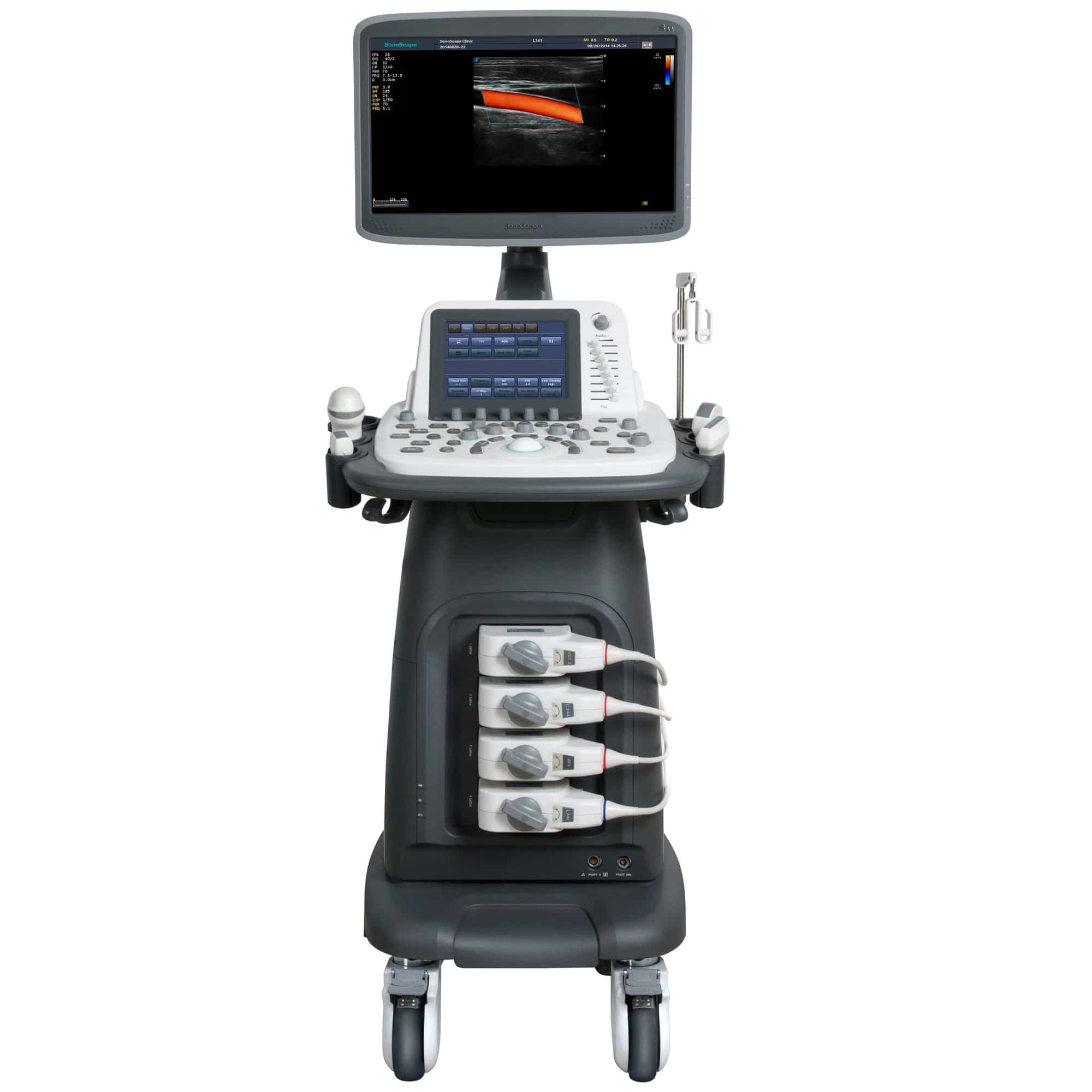
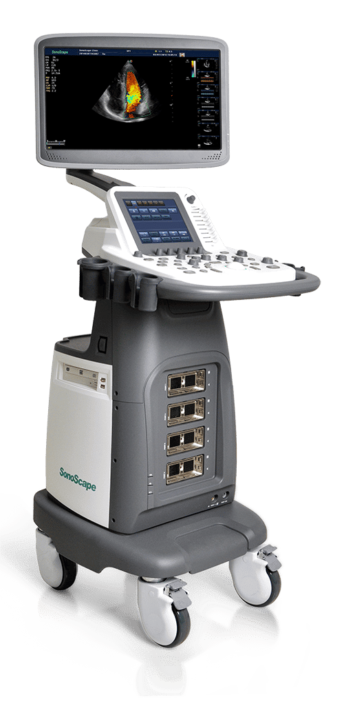
SonoScape S22
Excellence – mobile anywhere
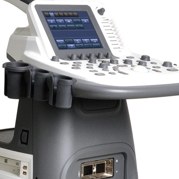
Display: 18.5″ wide screen LCD monitor, articulated arm, touch screen control panel
Weight: 65 kg
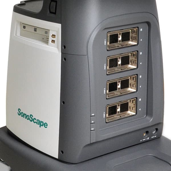
Probes: 4 probe ports (linear, convex, endo, biplane, TEE) + 1 pen probe
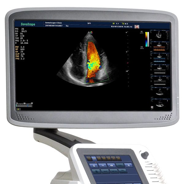
Imaging functions: B, 2B, M, THI, Color, DPI, PW, CW, TDI, 3D, 4D, Anatomical M-Mode, Stress Echo
Memory: internal on HDD
Software: DICOM 3.0, M-Tuning, measurement & calculation software, full digital beamformer, elastography, μ-Scan, panoramic imaging, ECG, auto. IMT
Measurement, simultaneous BiPlan
Connections: VGA, video out, 5x USB, S-Video, ECG, LAN, remote, DVD-RW, footswitch.
Please call today!
Description
SonoScape continues to go the extra mile to make its ultrasound technology even more valuable and efficient. The SonoScape S22 is designed to be a user-friendly platform that meets current and future challenges in women’s medical care. The ergonomic design and mobility of the SonoScape S22 not only benefits clinicians, but provides comfort to patients during exams.
Based on SonoScape’s proven technology, the SonoScape S22 brings you a new generation of superior image quality, especially in detecting abnormalities and small lesions – for more precision and consistency in diagnosis.
Outstanding performance
Phase Inversion Harmonic Imaging (PIH).
By emitting two ultrasound waves with opposite phase, PIH can equalize the fundamental and double the harmonic to reduce noise and spurious spots, and keep the harmonic at its maximum. This provides the user with a higher contrast image resolution that can visualize fine lesions, small structures, vessels, etc.
Compound Imaging
By deflecting the sound beams and using different frequency ranges, this technology superimposes the images. This provides optimal resolution, spurious pixel reduction and edge detection – for a clearer image and better continuity of structures, especially in surface and abdominal imaging.
µ-Scan
Our new-generation µ-Scan technology significantly improves the visibility of organs and lesions. High-definition contrast resolution suppresses spurious pixel artifacts while preserving true tissue structure.
Precise diagnostic results in obstetrics and gynecology
First class volume probe
Its ergonomic design allows the probe to move freely in the physician’s hand for efficient examination, yet is not uncomfortable for the patient during the examination.
Endocavitary probe with wide scanning angle
Combined with unique temperature sensor technology, the SonoScape S22 significantly shortens the exam – yet is more comfortable and safer for the patient.
Flexible key assignment by the user
With the exception of the professional obstetrics/gynecology measurement and related package, the SonoScape S22 is equipped with user-definable direct keys to make diagnosis more efficient.
Ultrasound guided puncture
Ultrasound diagnostics and interventions under ultrasound guidance are becoming more common. The SonoScape S22 offers you a range of biopsy guides for ultrasound-guided puncture for various clinical applications. Biopsy with the biplanar probe for implantation expands the range of applications, allowing not only ultrasound-guided punctures for diagnosis, but also enabling treatments.
Dual real-time imaging
This technology can display real-time B-mode and color mode simultaneously on the screen, allowing visualization of anatomy and blood flow during each examination. Real-time imaging is very convenient during exams because it means doctors don’t have to keep switching modes. Simply move the sonic probes and find the lesions.
Outstanding 3D/4D image quality
The special image quality in 3D/4D enables the best visualization of the fetus, providing doctors with so much information. This feature is particularly in demand in obstetrics. And numerous other application solutions, such as the multi-slice view and the measurement function, ensure that all ultrasound requirements are met – those of the mother and those of the physician.
Reliable scanning
The SonoScape S22 excels in radiology by combining its outstanding imaging technologies and unique probes with its unique hardware and software.
Excellent 2D image quality, even in color mode
Multi-beam, high-density, high-frequency probes and advanced ultrasound technologies help you detect even the smallest changes in anatomy and small structures.
Real-time panoramic imaging
Real-time panoramic imaging gives you a larger view – providing you with a larger comparative image for documenting the spatial relationship of structures in 2D and color Doppler modes.
High sensitivity for blood flow measurement
It easily acquires blood flow data, displays tiny vessels and outputs velocity data. This gives you a diagnostic image without overflow and with less background noise – for accurate diagnosis.
Trapezoidal imaging
By magnifying the image view with this technology, physicians get more data for faster diagnosis. With this feature, doctors can see more data at the same time.
© 2022 SonoMedical GmbH