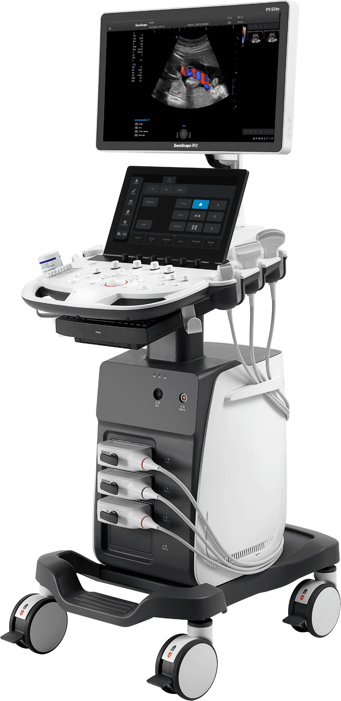
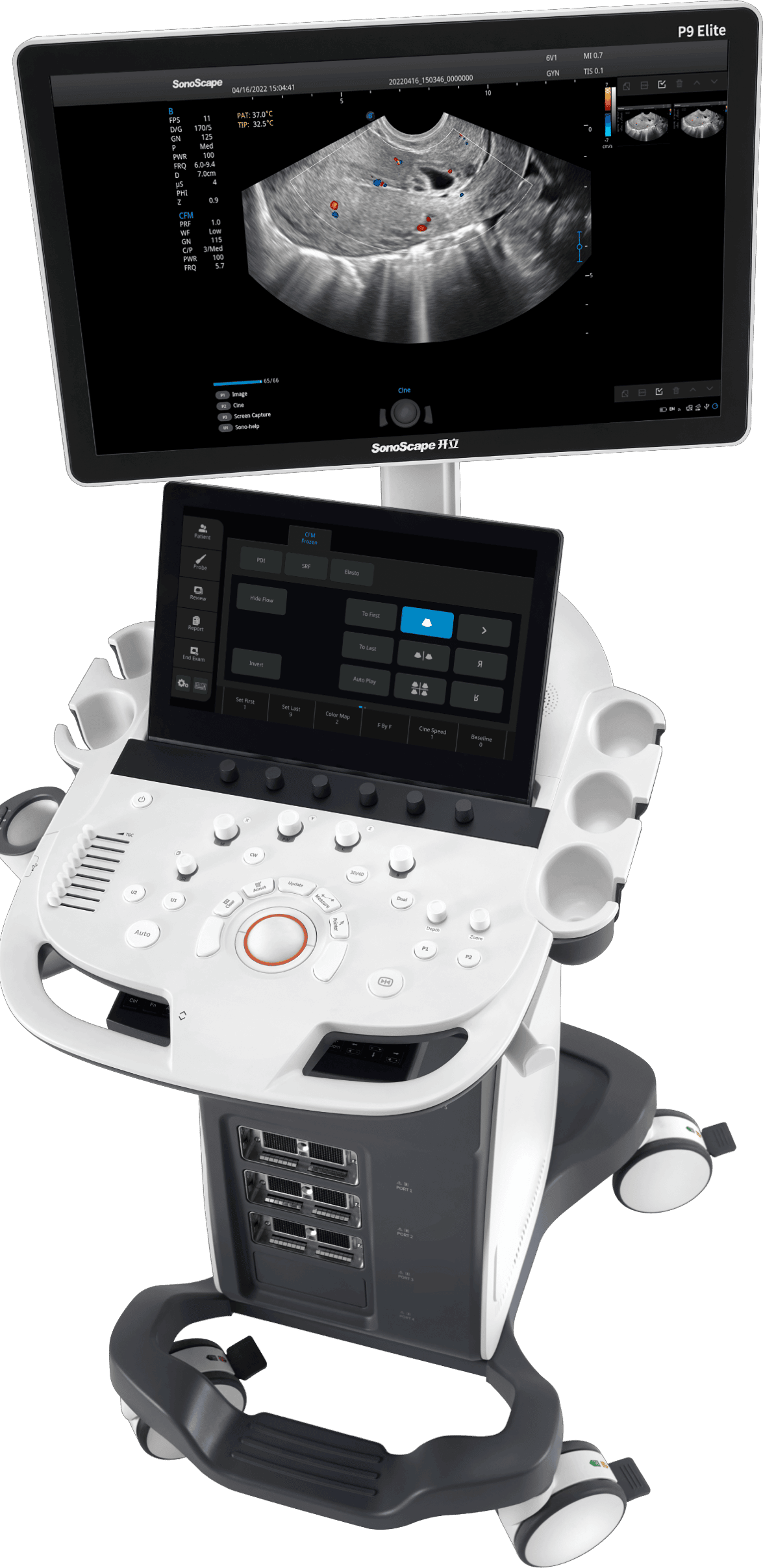
Devices Hardware:
- Flat panel monitor 21,5″ LED
- Scan image with extra large display in linear and endo areas
- "Single Crystal" transducers with C-field beam former
- Clearly arranged keyboard with additional 13.3″ touch screen for optimal operation
- Assignment of the preset program
- Very large cine memory
- Highly modern system architecture
- Small stand, tray for accessories and footrest are standard
- High mobility factor
Software:
- Latest image technology for excellent image quality
- User-friendly menu navigation
- Comprehensive measuring software for all disciplines
- THI Tissue Harmonic Image
- TDI (Tissue Doppler)
- Color Doppler (Dynamic Color Technology) Triplex, PW and CW
- Directional Power Doppler
- Trapezoidal display
- Preset Management
- Pulse Inversion
- Color Doppler Panorama
- Microscan Speckle Reduction
Image archiving:
- PC connection prepared
- Dual USB 3.0 DVD-RW
- DICOM and WORKLIST included at no extra charge
- Internal image storage on large hard disk
Please call today!
Description
The new engineered SonoScape P9-Elite color doppler ultrasound system is designed to acquire high-quality images, it features versatile probe configurations, multiple clinical tools, and automated analysis software to provide you with a comprehensive solution for your growing clinical application demand.
Classic Features
C-field beam
Unlike traditional focus, which concentrates on limited areas, the C-field beam, with its continuous dynamic focus, distributes signal energy evenly, contributing to a more uniform overall image.
Dynamic multi-beam technology
It is used to dynamically deliver multiple beams in different scan modes to balance parameter requirements in different applications. This provides the user with detailed data with good spatial resolution or real-time motion with suitable line density and frame rate.
Pure Inversion Harmonic Imaging
It fully preserves the harmonic signals without degrading the acoustic data. This improves the contrast resolution by reducing the noise and clutter of small structures, lesions, vessels, etc.
Spatial Compound Imaging
Spatial Compound Imaging utilizes multiple slice lines for resolution with optimal contrast, for spurious pixel reduction and edge detection. This feature of the Sonoscape P10 provides clearer images and shows continuous structures more clearly - making it an ideal system for superficial and abdominal imaging.
Special Functions
SR Flow
With the new innovative SR Flow technology, low velocity flow signals can be better detected. It also improves spatial resolution and also eliminates signal overflow to provide users with true hemodynamic data.
Vis-Needle
The Vis-Needle function is based on the steering and deflection of the ultrasound beam. It improves visualization of needle position in tissue to minimize potential damage to surrounding tissue, increase initial success rate, and reduce the risk of needle puncture.
Real-time panoramic imaging
Real-time panoramic imaging allows you to scan a larger field of view for large organs or lesions - for easy measurements and diagnostic efficiency.
WideScan
The WideScan feature magnifies the ultrasound image during a real-time scan with linear or convex probes for a complete view of large lesions or anatomical structures.
The SonoScape P10 offers a comprehensive selection of electronic probes that maximizes its usefulness for a variety of applications such as abdominal examination, pediatric, obstetrics and gynecology, cardiovascular, musculoskeletal, and more. Advanced probe technology also improves image quality and reliability in making clinical diagnoses - even in problematic patients.
Convex Probe 3C-A
Ideal for a wide variety of applications including abdomen, gynecology, obstetrics, urology and even abdominal biopsy.
Phase Array Probe 3P-A
For cardiology and emergency medicine examinations in adults and children, the phase array probe offers comprehensive presets for different examination modes - even for problematic patients.
Intracavitary Probe 6V1
The Intracavitary Probe can be used for gynecology and urology exams as well as prostate exams. Its temperature sensor technology not only protects the patient, but also extends its service life.
© 2022 SonoMedical GmbH