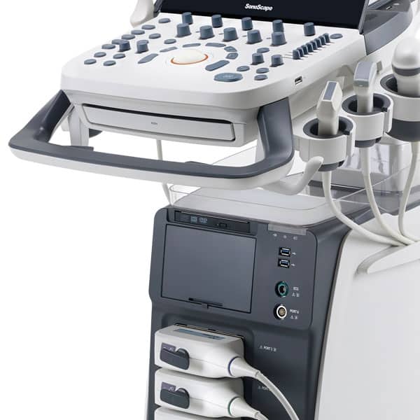
Devices Hardware:
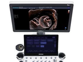
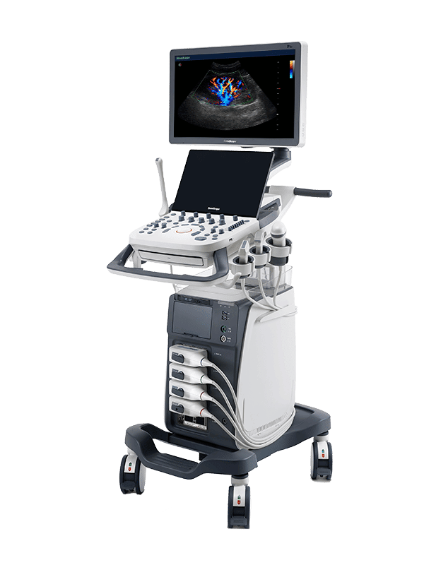
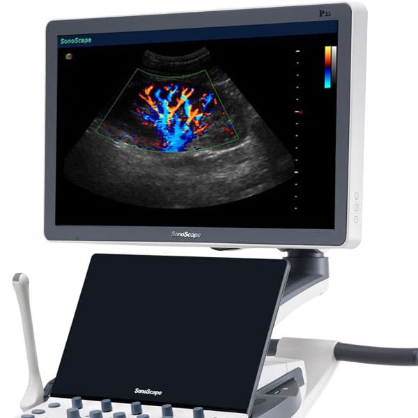
- 21.5 “high-resolution color LED monitor on multifunctional articulating arm
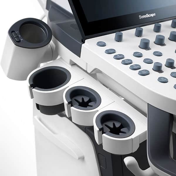
- Height-adjustable, rotatable, user-programmable control panel
- Additional 13.3″ touch screen
- Backlit, retractable keyboard
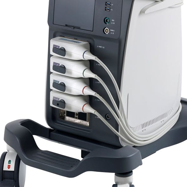
- Five probe slots (four active + one “parking”)
- “Single Crystal” peeling heads with C-field beam former
- Very large cine memory
- One pen probe port
- Wireless Wifi connection
- Internal memory on HDD
Software:
- Extensive measurement software for all disciplines
- 4 D image display
- 2D speckle reduction technology
- B (2B & 4B) mode
- Color Doppler flow imaging
- Pulse wave Doppler imaging
- HPRF
- Continuous Wave Doppler Imaging
- Power Doppler Imaging / Directional
- Power Doppler Imaging
- U-scan for Speckle Reduction
- Color Doppler (Dynamic Color Technology) Triplex, PW and CW
- Trapezoidal imaging
- Preset Management
- Auto NT
- Color Doppler Panorama
- M-mode
- Tissue harmonic imaging
- Tissue specific imaging
- Pulse Inversion Harmonic Imaging
- Spatial Imaging
- LGC (Lateral Gain Compensation)
- Full frame zoom
- Image Rotation
- Real-time 2D panoramic imaging- 4 D image display
- ECG Module
- THI Tissue Harmonic Image
- TDI (Tissue Doppler)
- Auto – IMT measurement
- Auto M-tuning, optimal image and Doppler settings at the touch of a button
Probes:
Linear:
- L741
- L742
- L752
Convex:
- 3C-A
- C1-6
- C613
Phased Array:
- S1-5
Endocavitary:
- 6V1
- 6V3
Volume:
- VC6-2
- (data are partly optional)
Please call today!
Description
P20’s user-friendly design with innovative technologies, a simple control panel, intuitive user interface and a variety of intelligent auxiliary scanning tools greatly enhances your daily exam experience. In addition to general imaging applications, P20 is entitled to diagnostic 4D technology that provides exceptional performance in obstetrical and gynecological applications.
Improved image quality with greater clarity
SonoScape continues to make strides in improving the image quality of its ultrasound products to increase physician confidence in diagnosis. With exceptional images from P20, anatomical structures are clearer than ever.
Value with compromise, treatment with confidence
C-xlasto Imaging
With C-xlasto Imaging, P20 enables comprehensive quantitative elastic analysis. Meanwhile, C-xlasto on P20 is supported by linear, convex and transvaginal probes to ensure good repeatability and highly consistent quantitative elastic results.
Contrast Imaging
Contrast imaging with 8 TIC curves enables clinicians to evaluate perfusion dynamics in a variety of clinical settings, including lesion location and assessment.
S-Live
S-Live provides detailed visualization of subtle anatomical features, enabling intuitive diagnosis with real-time 3D images and enriching patient communication.
Pelvic Floor 4D
Transperineal 4D pelvic floor ultrasound can provide useful clinical values for assessing the impact of vaginal delivery on the female anterior compartment by evaluating whether or not the pelvic organs are prolapsed and the extent to determine if the pelvic muscles have been accurately torn.
Anatomic M Mode
Anatomic M Mode helps you observe myocardial motion at different stages by freely placing sample lines. It accurately measures myocardial thickness and heart size of even difficult patients and assists in myocardial function and LV wall motion assessment.
Tissue Doppler Imaging
P20 is equipped with Tissue Doppler Imaging, which provides velocities and other clinical information on myocardial function, allowing clinical clinicians to analyze and compare the motion of different parts of the patient’s heart.
© 2022 SonoMedical GmbH